Have you ever stomped a spider and noticed a strange, milky-looking substance mixed in with the bits of bristly legs and carapace? Where is the bright red blood that we humans associate with injury? Do spiders even have blood?
Spider blood is called “hemolymph”. Hemolymph functions similarly to the blood found in humans and other vertebrate animals. But unlike blood, hemolymph washes freely through spiders’ bodies, fluoresces under ultraviolet light, helps spiders move, and is a bluish-green – rather than red – fluid.
Arthropods like spiders are fundamentally different from mammals like humans, cats, and dogs – even the fluid that flows through their circulatory systems to keep them alive is different. Read on to learn about spider blood and how it differs from the blood we are all used to.

Blood vs. hemolymph
“Blood” is the correct term for the fluid which flows through the circulatory systems of vertebrate animals like mammals.
“Hemolymph” is the correct term for the fluid which flows through arthropod systems. Arthropods are those animals classified in phylum Arthropoda and includes animals with segmented bodies, hard exoskeletons, and jointed legs, like spiders, insects, scorpions, crabs, and shrimp.
Function of hemolymph
Like humans, spiders are multi-cellular creatures whose body tissues are made up of thick layers of packed cells. Individual cells need a steady supply of nourishment, electrolytes, hormones, vitamins, and oxygen to metabolize energy, and also need metabolic waste products and carbon dioxide removed to avoid poisoning.
All animals also need to remain hydrated, fight off bacteria, viruses, and other invading microorganisms, and clot off wounds to survive the immediate aftermath of traumatic injury.
Blood supports these actions these actions in humans; hemolymph performs them in spiders.
Composition of hemolymph
Both hemolymph and blood are made up of water and a variety of specialized cells and chemicals. In both fluids, the water prevents dehydration and acts as a medium into which important nutrients and biochemicals dissolve for easy transport around the body. However, the types of cells and the chemicals dissolved in water are different in hemolymph than in blood.
Osmotic pressure
Osmotic pressure develops when two solutions of different concentrations are separated by a semipermeable membrane; osmotic pressure is higher on the side of the membrane with greater solute concentration, causing the solute to move to the side with lower pressure. In this way, important chemicals are transported into and out of cell membranes.
The osmotic pressure in most animals is created by inorganic ions such as sodium and chloride but in insects, organic molecules like free amino acids are more important (Barnes 1974).

Types of cells
Both vertebrate blood and spider hemolyph contain individual cells which perform specialized functions as they flow through the body but the type of cells in each are different.
A major component of vertebrate blood are cells called “erythrocytes”. Erythrocytes are mature red blood cells made in bone marrow and the average human has about five million blood cells per microliter of blood (Taber’s…1997). Each cells has an outer membrane and an internal framework made up of proteins and fats to which more than 200 million molecules of a special protein called “hemoglobin” attaches (Taber’s…1997).
Spiders have neither bones nor bone marrow tissue; their hemolymph does not contain erythrocytes.
Hemolymph contains cells called “hemocytes”, which are part of spiders’ immune systems. Hemocytes defend against infection by engulfing an invading microorganism and either destroying it outright in a process called “phagocytosis” or essentially walling it off from the rest of the spider’s body by forming a cyst or nodule (Resh and Carde 2009).
Vertebrate blood also contains phagocytic immune cells called neutrophils, monocytes, and macrophages, but these cells are different from the hemocytes found in spider hemolymph.
Oxygen-binding pigment
Another major difference between blood and hemolymph is how each carries oxygen.
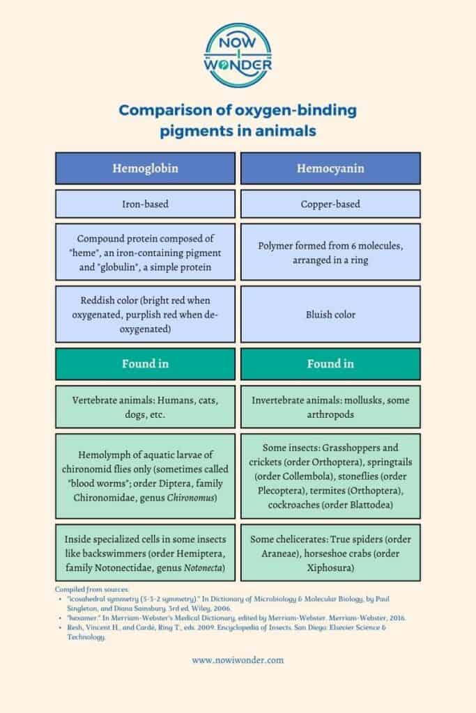
In vertebrate blood, oxygen is carried by the hemoglobin molecules found by the millions in each erythrocyte. Hemoglobin is a complex protein made of two parts: an iron-containing pigment called “hematin” and a protein called “globulin”. Oxygen is attracted to hemoglobin and binds to it readily to form a combination molecule called “oxyhemoglobin”. Oxyhemoglobin is an unstable compound and releases its oxygen to cells as the erythrocytes swirl through the blood stream.
In arthropod hemolymph, oxygen is carried by a completely different molecule called hemocyanin (Rehm et al. 2012). Unlike hemoglobin, which is iron-based and red, hemocyanin is copper-based and therefore bluish-green.
True spiders transport oxygen using hemocyanin. However, arachnids classified in taxon Apulmonata have lost their hemocyanin; these include harvestmen (order Opiliones), pseudoscorpions (order Pseudoscorpiones), wind scorpions (order Solifugae), and mites and ticks (order Acari) (Rehm et al. 2012).
Movement throughout the body
Spiders and other arthropods have open circulatory systems. Also called “lacunar” systems, these open circulatory systems are very different from the closed systems found in humans and other vertebrate animals.
Vertebrate circulatory systems are called “closed” because blood is fully contained within blood vessels (arteries, arterioles, capillaries, venules, and veins) during its passage through the body. Blood never leaks into the interstitial space (the area between cells) or mixes with extracellular fluid (fluid found outside cells) unless the blood vessel wall is injured, allowing the contents to escape.
Spiders – like all arthropods – have a totally different internal design. They have no veins and no physical separation between their hemolymph and their interstitial fluid (the fluid between individual cells).
Hemolymph leaves the heart through arteries but then washes freely into and through the body cavity, which is called a hemocoel. As it circulated through the hemocoel, the hemolymph releases oxygen, nutrients, hormones, electrolytes, etc. to the cells and picks up carbon dioxide and other waste products.
Eventually, hemolymph collects into spaces called “venous sinuses” and returns to the heart through tiny holes in the heart wall called “ostia” (Pechenik 2000).

Mechanical power
Spiders use their hemolymph to move their bodies; it transmits force generated by muscles through volume and pressure changes. This means of both generating movement and using circulatory system fluid is unique to true spiders. No other animal, including other arthropods, uses this method to move.
Movement in most animals is generated when two opposing muscles coordinate their contractions to shift the position and angle of a joint. For example, in humans, the biceps and triceps muscles are an opposing pair. When the biceps contracts, the triceps relaxes, and the forearm moves closer to the upper arm by rotating at the elbow joint. The elbow joint straightens when the biceps muscle relaxes and the triceps muscle contracts.
Spiders are a different story. They move by means of a unique “semi-hydraulic” system that relies on volume and pressure changes transmitted by their hemolymph.
Spiders have only one set of leg muscles – the flexor muscles, which bend the leg sections at the joints. They lack extensor muscles and extend their legs by contracting special muscles in their cephalothoraxes. When these muscles contract, hemolymphatic pressure around the leg joints increase and causes the joints to straighten (Brandt et al. 2021).
Fluorescence
Another unique aspect of spider hemolymph is that it fluoresces under short wavelengths of light (Brandt and Masta 2017).
Fluorescence is the “emission of a longer wavelength light by a material exposed to a shorter wavelength light” (Taber’s …1997). Spider hemolymph will glow under a “black light”, which is a special type of light bulb that radiates light in the short wave, ultraviolet portion of the electromagnetic spectrum.
Note that spider hemolymph is fluorescent only and does not glow in the dark; it glows only while exposed to the UV light. Objects that glow in the dark do so because they are phosphorescent rather than fluorescent and emit visible light even after the inducing radiation stops.
Conclusion
Spider hemolymph may not look very much like the bright red blood we’re all familiar with but it is as vitally important to spiders as blood is to us. Hemolymph serves many of the same functions as vertebrate blood but is different in many ways. Looks can be deceiving but one can’t be too surprised that the differences between a tarantula and a kitten are more than simply skin deep.
Related Now I Wonder Posts
For more information about spiders, check out these other Now I Wonder posts:
- What are wolf spiders?
- Jumping Spiders #1 – An Introduction
- Jumping Spiders #2 – A look at their incredible vision
- Jumping Spiders #3 – A detailed look at a special skill: Jumping
- Jumping Spiders #4 – As Predators
- Jumping Spiders #5 – As Prey
- Are spiders bugs?
- Do spiders have teeth?
For more information about spider relatives in class Arachnida, check out these other Now I Wonder posts:
References
Barnes RD. 1974. Invertebrate zoology. 3rd ed. Philadelphia (PA): W.B. Saunders Company.
Brandt EE, Masta SE. 2017. Females are the brighter sex: Differences in external fluorescence across sexes and life stages of a crab spider. PLoS One. 12(5).
Brandt EE, Yoshan S, Elias DO, Natasha M. 2021. Jump takeoff in a small jumping spider. Journal of Comparative Physiology. 207(2):153-64.
Hickman CP Jr., Roberts LS, Larson A, L’Anson H, Eisenhour DJ. 2006. Integrated principles of zoology. 13th ed. New York (NY): McGraw-Hill Companies.
Pechenik JA. 1996. Biology of the invertebrates. 4th ed. New York (NY): The McGraw-Hill Companies, Inc.
Resh, Vincent H., and Cardé, Ring T., eds. 2009. Encyclopedia of Insects. San Diego: Elsevier Science & Technology.
Rehm P, Pick C, Borner J, Markl J, Burmester T. 2012. The diversity and evolution of chelicerate hemocyanins. BMC Evolutionary Biology. 12:19.
Taber’s Cyclopedic Medical Dictionary. 1997. 19th ed. Philadelphia (PA): F. A. Davis Company.